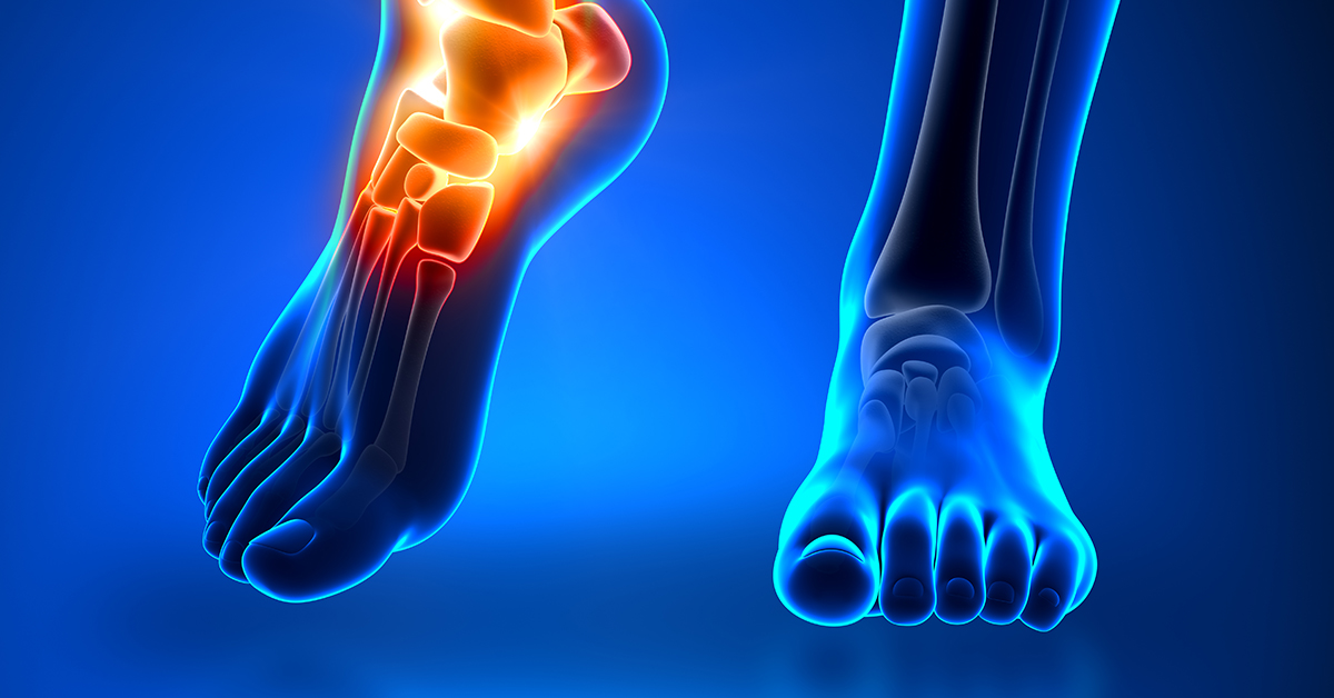
The Bones in the Foot
There are normally 26 bones in the foot. These bones are divided into three different groups: tarsal bones, metatarsal bones, and phalanges. Each of the bones in the foot has one or more articulating surfaces, meaning they form a joint with another bone, resulting in more than 30 joints in the foot. In addition to those 26 articulating bones, there are also two sesamoid bones, which are bones that do not attach to any other bone and instead serve to help the functioning of muscles and ligaments.
Hindfoot
The tibia and the fibula, the two lower leg bones, form the ankle joint with the talus, a small tarsal bone located between lower leg bones and the calcaneus, or heel bone. The calcaneus is the largest of the tarsal bones, and it forms the heel of the foot and bears the body's weight.
Midfoot
Two of the seven tarsal bones are in the hindfoot, while the other five tarsal bones form the midfoot and help build the foot's arch. In order from the hindfoot forward, these tarsal bones are the navicular, cuboid, the medial cuneiform, the middle cuneiform, and the lateral cuneiform. The heads of the cuneiform bones meet and articulate with the bases of the metatarsals, which is where the forefoot begins.
Forefoot
The forefoot is composed of five metatarsal bones and the 14 phalanges. The metatarsal bones are numbered from one through five, starting with the metatarsal on the medial side, which is the side your big toe is on. Each of the metatarsals has a base, a shaft, and a head. Each metatarsal's base articulates with one or more tarsal bones while the head of each metatarsal articulates with the base of the phalanges. The two sesamoid bones can be found under the first metatarsal head.
The phalanges are the bones in your toes. Each of your smaller toes has 3 articulating phalanges. From the base of the toes to the end, these bones are the proximal phalange, the middle phalange, and the distal phalange. The big toe is the exception to this, having only a proximal phalange and a distal phalange.
Soft Tissue in the Foot
Many different tendons, ligaments, and muscles work together with the bones in the foot to make movement, balance, weight-bearing, and other tasks pain-free and easy. These tissues also help the foot maintain its structure, with ligaments connecting bone to bone and tendons connecting muscle to bone. So issues with any structure in the foot can be very painful. For example, one of the foot's ligaments, called the plantar fascia, supports your foot's arch while connecting the heel to the front of the foot. This ligament absorbs strain and stress exerted on the foot, but like any tissue, the plantar fascia can be damaged if it's overloaded or overused. This damage prevents it from fulfilling its role as it normally would, which can result in pain and other issues.
